
At-home melanoma detection? Microneedle skin patch under development
- Allan Ryan
- Aug 11, 2025
- 2 min read

A team at the University of Michigan has developed a microneedle skin patch that can detect melanoma by sampling biomarkers in the epidermis, offering a possible pathway to at-home testing for this aggressive form of skin cancer.
The device, called the ExoPatch, uses star-shaped silicone microneedles measuring 0.6 mm in length and less than 100 nanometers at the tip. “The star-shaped needles make puncture easier and less painful, but they are so small that they only go through the top-most layer of the skin, the epidermis, and do not draw blood,” said Sunitha Nagrath, PhD, the Dwight F. Benton Professor of Chemical Engineering at U-M and co-corresponding author of the study published in Biosensors and Bioelectronics.
Unlike current melanoma screening methods that often require biopsies and waiting periods for pathological assessment, the ExoPatch is applied for 15 minutes, then dissolved in acid to release captured exosomes—the cellular messengers often elevated in cancer. “A fair-skinned person with moles must go to the doctor about every six months for a biopsy to see if they’re malignant or benign. With this test, they could instead test at home, get the results right away and follow up with a dermatologist for a positive result,” Dr. Nagrath said in a press release.
The exosomes are detected by a simple lateral flow immunoassay in which two lines signify the presence of melanoma markers and one line indicates a negative result, echoing the format of at-home Covid-19 tests. In preclinical studies, the ExoPatch successfully distinguished melanoma tissue from healthy tissue in mice, producing a test strip line 3.5 times darker in melanoma-positive samples. Quantitative analysis showed that the patch isolated 11.5 times more exosomal protein from melanoma tissues compared to healthy controls, indicating the device’s selectivity for cancer-specific exosomes.
“When looking at microscopy images, I was happy to see how nicely the exosomes adhered to the microneedles and were within the 30 to 150 nanometer size range we expect,” said Scott Smith, a doctoral student and co-lead author of the study.
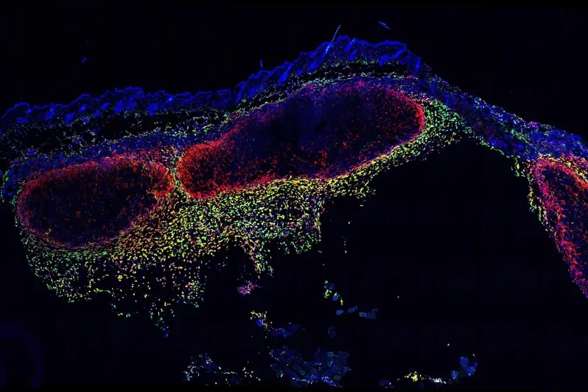
Pig skin, chosen for its close similarity to human skin in thickness and composition, was used to validate epidermal penetration. Next steps include pilot studies in humans and subsequent clinical trials to assess safety and accuracy. The research team notes that the ExoPatch gel could be adapted to detect exosomes released by other solid tumours, potentially broadening the application to other cancers such as lung, breast, colon, prostate, and brain.
“This is the first patch designed to capture disease-specific exosomes from fluid under the skin. The potential applications are huge,” Dr. Nagrath said.

Comments