
Some skin cancers may start in hair follicles
- By Dhiren Mahiban
- Nov 4, 2019
- 3 min read

Researchers at the NYU School of Medicine and Perlmutter Cancer Center have found that the most deadly skin cancers may start in stem cells that lend colour to hair, and originate in hair follicles.
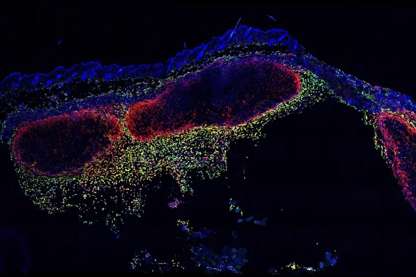
The study published online ahead of print in Nature Communications (Nov. 4, 2019) found that unlike their normal counterparts, newly cancerous pigment stem cells migrate up and out of the follicles to establish melanomas in nearby surface skin before spreading deeper. The study was conducted in genetically engineered mice, with the results confirmed in human tissue samples.
“By confirming that oncogenic pigment cells in hair follicles are a bona fide source of melanoma, we have a better understanding of this cancer’s biology and new ideas about how to counter it,” said study author, Dr. Mayumi Ito Suzuki, in a press release. Dr. Suzuki is an associate professor in the Ronald O. Perelman Department of Dermatology at NYU School of Medicine and Perlmutter Cancer Center.
Past models of the disease suggested that sunlight was a major risk factor for melanoma, but current research argues that the triggers are always there in normal follicles.
The new study looks at the stem cells that mature into melanocytes, cells that make the protein pigment melanin, which protects skin by absorbing some of the sun’s ultraviolet, DNA-damaging rays. By absorbing some wavelengths of visible light, but reflecting others, pigments create hair colour.
In a series of steps, the NYU research team established a new mouse model for the study of melanoma, so that the team could edit genes in follicular melanocyte stem cells only (the c-Kit-CreER mouse). This capability enabled researchers to introduce genetic changes that made only melanocyte stem cells, and their descendants destined to form melanomas, glow no matter where they travelled.
With the ability to accurately track a key stem cell type for the first time, the study’s authors confirmed that melanoma cells can arise from melanocyte stem cells, which abnormally migrate up and out of hair follicles to enter the epidermis. The team then tracked the same cells as they multiplied there, and then moved deeper into the skin layer.
Once in the dermis, the cells shed the markers and pigment that went with their follicular origins, presumably in response to local signals. They also acquired signatures similar to nerve cells (neurons) and skin cells (mesenchymal), molecular characteristics similar to those noted in examinations of human melanoma tissue.
Knowing where to look for the original, cancer-causing event, the researchers temporarily eliminated signals one by one in the follicular environment to see if cancer still formed in their absences.
As a result, the researchers confirmed that follicular melanocyte stem cells, even though they had cancer-causing genetic mutations, did not multiply or migrate to cause melanomas unless also exposed to endothelin (EDN) and WNT. These signalling proteins normally cause hairs to become longer and pigment cells to multiply in follicles.
“Our mouse model is the first to demonstrate that follicular oncogenic melanocyte stem cells can establish melanomas, which promises to make it useful in identifying new diagnostics and treatments for melanoma,” said Dr. Qi Sun, a member of the research team. “While our findings will require confirmation in further human testing, they argue that melanoma can arise in pigment stem cells originating both in follicles and in skin layers, such that some melanomas have multiple stem cells of origin.”
Comments