
New tracer may improve PET scan detection of melanoma
- by John Evans
- Jan 18, 2019
- 2 min read
A new tracer molecule for using positron emission tomography (PET) imaging to detect malignant melanoma has been successfully tested in humans and could represent a way to improve detection of primary and metastatic melanoma.
These findings were published in The Journal of Nuclear Medicine (Jan. 1, 2019; 60(1):16–22).
The technique involves using a new tracer molecule, N-(2-(diethylamino)-ethyl)-18F-5-fluoropicolinamide (18F-P3BZA), which targets melanin pigment.
“While melanin-targeted PET probes have been studied in small animal models for a long time, this is the first time such a probe has been successfully translated into clinical evaluation,” the paper’s senior author Zhen Cheng, PhD, explained in a press release. “Our research shows that 18F-P3BZA is safe and, moreover, it is highly promising for clinical diagnosis and staging of melanoma.”
Dr. Cheng is an associate professor of radiology, and a member of the Molecular Imaging Program at Stanford, Bio-X Program, Stanford Cancer Center and Canary Center at Stanford for Early Cancer Detection, in Palo Alto, Calif. He is also the director of Stanford’s Cancer Molecular Imaging Chemistry Laboratory (CMICL).
To test the new molecular tracer, six healthy individuals were injected with 18F-P3BZA and then received whole-body PET/CT scans and blood tests to assess biodistribution, pharmacokinetic, and radiation dosimetry at 10 minutes, one hour, two hours, and four hours post-injection.
As well, five patients with suspected melanomas received 18F-P3BZA PET/CT imaging at 10 minutes and 1 hour after injection. For comparison of diagnostic ability, all patients also received 18F-fluoro-2-deoxyglucose (18F-FDG) PET/CT scans on the third day.
The investigators found that 18F-P3BZA was safe and clearly delineated melanoma tumours in patients.
“Given its specific melanoma-imaging capability, 18F-P3BZA is expected to be a new probe which may overcome some of the limitations of 18F-FDG, such as the false positive of inflammation and a substantially lower tumour-to-muscle contrast compared with 18F-P3BZA,” said Dr. Cheng. “Therefore, 18F-P3BZA may improve melanoma and metastasis detection and help guide therapeutic planning for melanoma patients.”
Dr. Cheng noted: “Our findings highlight the promise and importance of developing novel molecular probes for cancer imaging and therapy guidance.”

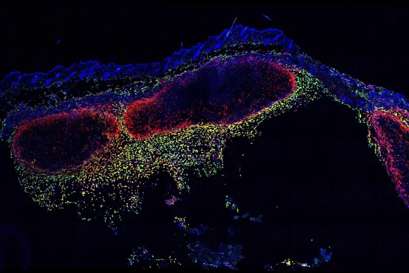
Sample images of a melanoma patient. (A) Maximum-intensity-projection PET images at 10 and 60 minutes after 18F-P3BZA injection and at 60 minutes after 18F-FDG injection. (B) Transverse images of primary melanoma (red arrow), lymph node metastasis (yellow arrow), and bone metastasis (blue arrow). (C) H&E (upper part) and immunohistochemistry (lower part) images of primary melanoma. Photo by: Xiaowei Ma, Shengjun Wang et al., Xijing Hospital, Fourth Medical University, Xi’an, China, and Stanford University, Stanford, CA.
Comments